Table of Contents
What is superresolution microscopy?
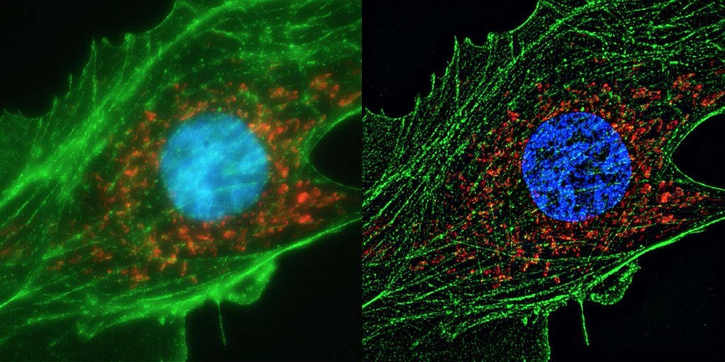
University of Michigan researchers have created a ground-breaking superresolution method that enables in-depth imaging of cell division at the nanoscale. This revolutionary technique has created new opportunities in the field of biology because it does not rely on molecules that break down over time. Superresolution microscopy, a method that can detect objects as fine as 10 nanometers, or roughly the breadth of 100 atoms, has transformed biological study since its invention and was awarded the Nobel Prize in 2014.
For ages, microscopy has been a pillar of scientific inquiry because it enables us to see objects that are invisible to the human eye. The zooming limitation placed a ceiling on the resolution of conventional light microscopes, restricting the detection of structures smaller than roughly half the wavelength of light utilized for imaging. The development of superresolution microscopy methods was prompted by this constraint.
The invention of superresolution microscopy has triggered a revolution in the field of microscopy, where researchers seek to understand the complex structures and functions of cells and molecules. With the use of this cutting-edge technique, scientists can now see the nanoscale world with clarity that is unmatched by conventional microscopy. Recent discoveries made possible by superresolution microscopy have expanded the frontiers of knowledge and significantly altered our understanding of biology, medicine, and materials science.

How superresolution microscopy works?
Superresolution microscopy can be achieved by a range of techniques that achieve resolutions well beyond the diffraction limit. Some prominent methods include:
- Stimulated Emission Depletion (STED): STED microscopy uses laser pulses to deplete the fluorescence emission of molecules surrounding the focal point, allowing the precise localization of individual molecules and enabling resolutions of a few tens of nanometers.
- Structured Illumination Microscopy (SIM): SIM employs patterned light illumination to create higher-frequency spatial information that can be processed to enhance resolution. It achieves two-fold improvement over the diffraction limit.
- Single-Molecule Localization Microscopy (SMLM): Techniques like PALM (Photoactivated Localization Microscopy) and STORM (Stochastic Optical Reconstruction Microscopy) exploit the precise localization of individual fluorescent molecules to create high-resolution images.
- Expansion Microscopy: This innovative technique involves physically expanding the sample, allowing structures initially too close to be resolved. After expansion, the sample is imaged with a standard microscope.
Applications of superresolution microscopy:
Superresolution microscopy has led to numerous groundbreaking findings across various scientific disciplines:
- Cell Biology: Researchers have used superresolution microscopy to visualize cellular structures like synapses, organelles, and the cytoskeleton in unprecedented detail. This has uncovered new insights into cellular processes, including endocytosis, cell division, and organelle dynamics.
- Neuroscience: In neuroscience, superresolution microscopy has enabled the visualization of individual neurotransmitter vesicles at synapses and the fine details of dendritic spines, contributing to our understanding of neural communication.
- Cancer Research: Superresolution microscopy has illuminated the nanoscale organization of proteins within cancer cells, offering insights into tumor formation, migration, and response to treatment.
- Materials Science: Superresolution techniques have been applied to study nanomaterials, allowing researchers to analyze their morphology and interactions at the nanoscale. This has implications for designing more efficient energy materials and catalysts.
- Virology: Superresolution microscopy has revealed the intricate architecture of viruses, helping scientists understand their assembly, replication, and interactions with host cells.
In summary, superresolution microscopy is a game-changing technique that has broken down the limitations of conventional microscopy. With the help of its most recent discoveries, we can now view and understand the complexities of the nanoscale world, opening up a new field of scientific inquiry. Superresolution microscopy has the potential to solve more riddles, alter scientific paradigms, and spur innovation across a wide range of fields, ultimately improving our understanding of the fundamental processes that underlie life and matter.
